In wake of Trump’s fetal tissue clampdown, scientists strain to adjust

Renewals of two diabetes grants held by Megan Sykes (right) of Columbia University could be held up by a new federal policy scrutinizing fetal tissue research.Michael DiVito

By Meredith WadmanJocelyn KaiserJun. 11, 2019 , 3:00 PM

Megan Sykes, an immunologist at Columbia University, has spent years using human fetal tissue to develop a mouse with a humanlike immune system, which mimics how type 1 diabetes develops in humans. The tissue is donated after elective abortions, and the mice are testbeds for potential diabetes treatments. But last week, she learned that President Donald Trump, acting on a priority of advocacy groups opposed to abortion, had issued a new policy that is likely to cause lengthy delays the next time she seeks U.S. government grants for her work—and could even choke off federal funding for all studies that use fetal tissue. The policy “is incredibly disappointing,” Sykes says, because it is a “politically motivated decree” that could derail numerous disease research efforts.

The new Trump policy, issued 5 June after a 9-month review led by officials at the Department of Health and Human Services (HHS), has three major components. One kills a long-standing contract between the National Institutes of Health (NIH) in Bethesda, Maryland, and the University of California (UC), San Francisco, under which the university used fetal tissue to develop humanized mice for HIV drug testing. Another ends research using fetal tissue conducted by any scientist directly employed by NIH. The third and widest-reaching provision adds a lengthy and uncertain step to NIH’s process for awarding new or renewal grants to university scientists, such as Sykes, for studies that use human fetal tissue. It requires HHS to appoint a separate 14- to 20-member ethics advisory board to review each proposal that NIH reviewers have found worthy of funding. The review of up to 6 months will result in a funding recommendation to the HHS secretary, who can accept or reject the advice.

Enacting the new policy “was the president’s decision …. to protect the dignity of human life,” Judd Deere, deputy White House press secretary, told Science. It was applauded by antiabortion activists, whose lobbying prompted HHS to launch its review of U.S.-funded fetal tissue research in September 2018. “This is a major pro-life victory,” said Marjorie Dannenfelser, president of the Susan B. Anthony List in Washington, D.C.

Many biomedical researchers were stunned, noting that the tissue, which would otherwise be discarded, has properties that make it valuable for research. It is less specialized than adult tissue, for instance, and readily adapts to new environments. “These new restrictions have no scientific or ethical basis and will roll back decades of consensus in the U.S., delaying the development of new treatments,” said Doug Melton, president of the International Society for Stem Cell Research in Skokie, Illinois, and co-director of the Harvard Stem Cell Institute.

“The whole point here is to so wrap the research in red tape that it’s impossible or at least unlikely to be feasible for many researchers,” says bioethicist Alta Charo of the University of Wisconsin in Madison.

A 1993 law formalized rules for using fetal tissue donated after elective abortions in U.S.-funded research. Last year, NIH spent $115 million on roughly 173 projects that rely on the tissue; about 160 were run by university scientists. One-third of the 173 grants focus on HIV/AIDS, many using humanized mice to probe, for example, how HIV hides out and evades the immune system, and what drugs might defeat it. Others tackle other infectious diseases, eye disease, and fetal development as well as toxic exposures during pregnancy.

NIH says its scientists are conducting just three projects affected by the new rules; all will stop. “This decision is devastating. It effectively ends our studies looking into new approaches for an HIV cure,” says Warner Greene, director of the Gladstone Institutes Center for HIV Cure Research in San Francisco. Greene is a partner in one of the projects, run by retrovirologist Kim Hasenkrug of NIH’s Rocky Mountain Laboratories in Hamilton, Montana.

At universities, the policy allows existing projects to continue until their current NIH funding expires. Nearly half of these extramural grants will expire within the next 18 months, and scientists will need to apply for a renewal if they want to keep the work going. Grantees are now grappling with what the new review process might mean.

It has already caused at least one researcher to change course. HIV scientist Jerome Zack last week told colleagues at UC Los Angeles (UCLA) that he had decided to remove his work using fetal tissue to develop humanized mice from a renewal application, due at NIH in August, for a large grant supporting the university’s long-standing Center for AIDS Research. “The grant covers way more than mouse work, it covers all HIV research on campus,” he says. “I don’t want to jeopardize that.”

Scott Kitchen, a Zack collaborator who directs mouse production at UCLA, says that in the past year his group provided humanized mice for more than 70 scientists on campus and nine at other institutions, as well as performing multiple large projects for several companies. “All of this has been critical in scientific and therapeutic development,” Kitchen says. “And all of it may now be derailed.”

At Columbia, Sykes is worried about the one-third of her 15 staff who are funded through two NIH grants. She recently submitted a renewal proposal for one grant and planned to submit the other in July. HHS hasn’t said when the policy will kick in. But when Sykes asked NIH officials how it might affect her proposals, the response “wasn’t reassuring,” she says.

Much could depend on whom the HHS secretary appoints to the ethics review boards. Under existing law governing HHS ethics boards, one-third to one-half of a board’s members must be scientists, and each must include at least one theologian, one ethicist, one physician, and one attorney. The law “absolutely” would allow HHS to pack the boards with members who oppose abortion, Charo says.

Critics of the new policy also say it will undermine a goal of opponents of fetal tissue research: to find and encourage the use of alternatives. In December 2018, NIH Director Francis Collins noted that his agency was putting up to $20 million over 2 years into research on such alternatives. But scientists say that those alternatives need to be tested for validity against human fetal tissue itself. For the time being, Collins said in December 2018, fetal tissue would “continue to be the mainstay” for certain kinds of research.

The new rules could remove that mainstay. But Charo notes a new president could reverse the policy, which is not codified in law. 

Fonte/Source: https://www.sciencemag.org/news/2019/06/wake-trumps-fetal-tissue-clampdown-scientists-strain-adjust

According to ABCNews:

The Trump administration on Wednesday announced it is suspending the use of fetal tissue in research conducted by government scientists and said it is ending a contract with a California university over its use of the materials.

“Promoting the dignity of human life from conception to natural death is one of the very top priorities of President Trump’s administration,” said the announcement from the Department of Health and Human Services.

According to the Congressional Research Service, the use of fetal tissue for research in the United States dates back to the 1930s and has been used by the National Institutes of Health since the 1950s. Among a variety of medical research uses, fetal tissue is obtained through elective abortions and has been used to help develop vaccines against diseases like measles and polio, the CRS said.

University of California, San Francisco, Chancellor Sam Hawgood called the move “politically motivated, shortsighted and not based on sound science,” and noted that it ended a “30-year partnership with the NIH to use specially designed models that could be developed only through the use of fetal tissue to find a cure for HIV.”

“UCSF strongly opposes today’s abrupt decision by the Health and Human Services Department (HHS) to discontinue intramural fetal tissue research by scientists at the National Institutes of Health (NIH),” he said in a statement. “The efforts by the administration to impede this work will undermine scientific discovery and the ability to find effective treatments for serious and life-threatening disease.”(MORE: Trump administration scales back on migrant child education as apprehensions soar)

The announcement was applauded by anti-abortion advocates, including Susan B. Anthony’s List President Marjorie Dannenfelser, who called it a “major pro-life victory.”

“President Trump knows we can do better as a nation and we are encouraged to see NIH Director Francis Collins carry out the President’s pro-life commitment,” Dannenfelser said.

This move follows a review conducted by the Department of Health and Human Services which looked at “all HHS research involving human fetal tissue from elective abortions,” according to the announcement.

PHOTO: The US Department of Health and Human Services building in Washington, DC, July 21, 2007.
The US Department of Health and Human Services building in Washington, DC, July 21, 2007.

As a result of the review, the administration is letting its contract with the University of California, San Francisco expire on Wednesday. The contract was for “research involving human fetal tissue from elective abortions,” the agency said.

“The audit and review helped inform the policy process that led to the administration’s decision to let the contract with UCSF expire and to discontinue intramural research – research conducted within the National Institutes of Health (NIH) – involving the use of human fetal tissue from elective abortion,” HHS said.(MORE: Housing Secretary Ben Carson defends hot-button policies, laments ‘gotcha’ politics)

Last year, HHS said it was giving $20 million to research alternative methods to using fetal tissue in its research and, according to the agency, HHS will continue to do so, saying that they are “committed to providing additional funding to support the development and validation of alternative models.”

ABC News reported last year that the move to eliminate the use of fetal tissue in research could set up a potential clash between the White House and House Democrats, who said at the time they weren’t convinced there was an alternative to fetal tissue that would suffice.

On Wednesday, the chaiman of the House Health subcommittee, Rep. Anna Eshoo, D-Calif., said in a statement that the decision “puts politics over progress.”

“Research using fetal tissue has led to numerous vaccines and treatments that have saved millions of lives,” Eshoo said. “The Administration’s move to cut federal research, including the cancelation of UCSF’s HIV research contract, jeopardizes new cures for patients. This backward decision is another blow against science from the Trump Administration.”

ABC News’ Anne Flaherty contributed to this report.

Artificial lenses, heart valves, hearts, and (for the first time) pancreases

Top Left: Intraocular lens; Top Right: Artificial heart valve; Bottom Left: Artificial Heart; Bottom Right: Artificial Pancreas.

The motivation behind writing this article is that the U.S. FDA, for the first time, has approved an artificial pancreas. Artificial body parts which: see for us (intraocular lenses); allow our blood to flow in our hearts in a desired direction without backflow (artificial heart valves); pump our blood for us on the order of 7 liters per minute; and control the glucose level within our systemic circulation (which also controls how much glucose is within our cells). Startingly, we have approximately 1 trillion cells per kilogram of body mass. We have a lot of cells to take care of, given the average person weighs approximately 70 kg.

Biomedical engineering is transformative, to say the least. This article will briefly discuss discuss artificial lenses for eyes (intraocular lenses), artificial heart valves, hearts, and pancreata (pancreases).

Introacular lenses

Intraocular lenses used to be manufactured using poly(methyl methacrylate) or PMMA. PMMA was the material of choice because of the serendipitous observation made by “British ophthalmologist Sir Harold Ridley observed that Royal Air Force pilots who sustained eye injuries during World War II involving PMMA windshield material did not show any rejection or foreign body reaction. Deducing that the transparent material was inert and useful for implantation in the eye, Ridley designed and implanted the first intraocular lens in a human eye.” (-wikipedia).

Acrylic MICS-IOL in holder

Artificial heart valves

An artificial heart valve is a device implanted in the heart of a patient with valvular heart disease. When one of the four heart valvesmalfunctions, the medical choice may be to replace the natural valve with an artificial valve. This requires open-heart surgeryHeart valvesare integral to the normal physiological functioning of the humanheart. Natural heart valves induce unidirectional blood flow through the valve structure from one chamber of the heart to another. Natural heart valves become dysfunctional for a variety of pathological causes, some of which may require complete surgical replacement of the natural heart valve with an artificial valve. (-wikipedia)

Top: Starr-Edwards Valve
Middle: Starr-Edwards Valve
Bottom: Smeloff-Cutter Valve

Artificial hearts

An artificial heart is a device that replaces the heart. Artificial hearts are typically used to bridge the time to heart transplantation, or to permanently replace the heart in case heart transplantation is impossible. Although other similar inventions preceded it from the late 1940s, the first artificial heart to be successfully implanted in a human was the Jarvik-7 in 1982, designed by a team including Willem Johan Kolff and Robert Jarvik.

An artificial heart is distinct from a ventricular assist device (VAD) designed to support a failing heart. It is also distinct from a cardiopulmonary bypass machine, which is an external device used to provide the functions of both the heart and lungs and are used only for a few hours at a time, most commonly during cardiac surgery.

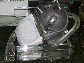
Artificial-heart-london.JPG
An artificial heart displayed at the London Science Museum

Origins of artificial hearts

A synthetic replacement for the heart remains a long-sought “holy grail” of modern medicine. The obvious benefit of a functional artificial heart would be to lower the need for heart transplants, because the demand for organs always greatly exceeds supply (rather necessary for transplants are normally unfit for transfer).

Although the heart is conceptually a pump, it embodies subtleties that defy straightforward emulation with synthetic materials and power supplies. Consequences of these issues include severe foreign-body rejection and external batteries that limit mobility. These complications limited the lifespan of early human recipients to hours or days.

Early development of artificial hearts

The first artificial heart was made by the Soviet scientist Vladimir Demikhov in 1937. It was transplanted to a dog.

On 2 July 1952, 41-year-old Henry Opitek, suffering from shortness of breath, made medical history at Harper University Hospital at Wayne State University in Michigan. The Dodrill-GMR heart machine, considered to be the first operational mechanical heart, was successfully used while performing heart surgery.[1][2] Ongoing research was done on young male cows at Hershey Medical Center, Animal Research Facility in Hershey, PA during the 1970’s.

Forest Dewey Dodrill, working closely with Matthew Dudley, used the machine in 1952 to bypass Henry Opitek’s left ventricle for 50 minutes while he opened the patient’s left atrium and worked to repair the mitral valve. In Dodrill’s post-operative report, he notes, “To our knowledge, this is the first instance of survival of a patient when a mechanicaly heart mechanism was used to take over the complete body function of maintaining the blood supply of the body while the heart was open and operated on.”[3]

heart–lung machine was first used in 1953 during a successful open heart surgery. John Heysham Gibbon, the inventor of the machine, performed the operation and developed the heart–lung substitute himself.

Following these advances, scientific interest for the development of a solution for heart disease developed in numerous research groups worldwide.

First U.S. FDA Approved Artificial Pancreas (Medtronic)

Picture of the device.

This is a brief overview of information related to FDA’s approval to market this product. See the links below to the Summary of Safety and Effectiveness Data (SSED) and product labeling for more complete information on this product, its indications for use, and the basis for the FDA’s approval.

Product Name: The 670G System
PMA Applicant: Medtronic MiniMed, Inc.
Address: 18000 Devonshire Street, Northridge, CA, 91325
Approval Date: September 28, 2016
Approval Letter: http://www.accessdata.fda.gov/cdrh_docs/pdf16/P160017a.pdf

What is it? The Medtronic MiniMed 670G System is the first FDA approved hybrid closed loop system that monitors glucose and automatically adjusts the delivery of long acting or basal insulin based on the user’s glucose reading.

How does it work? The Medtronic MiniMed 670G System consists of a continuous glucose monitor (CGM) that measures the user’s glucose levels for up to seven days, an insulin pump that delivers insulin to the user, and a glucose meter used to calibrate the CGM.

The MiniMed 670G System is able to decrease or stop insulin delivery when it detects the user’s glucose is low, or increase the insulin delivery when the system detects the user’s glucose levels are high with no input from the user. The glucose sensor contains a wire that is inserted under the skin on the abdomen and measures glucose values in the tissue fluid. The glucose values are wirelessly sent to the insulin pump, and displayed along with glucose trend information, alerts, and alarms on the pump screen. The insulin pump delivers a prescribed dosage of insulin through an infusion set. The insulin pump can automatically adjust the delivery of insulin using a mathematical equation, or algorithm that incorporates information from the CGM.

The system has two modes; Manual Mode and Auto Mode. While in Manual Mode, the system can be programmed by the user to deliver basal insulin at a preprogrammed constant rate. The system will automatically suspend delivery of insulin if the sensor glucose value falls below or is predicted to fall below a predetermined threshold. The system will also automatically resume delivery of insulin once sensor glucose values rise above or are predicted to rise above a predetermined threshold. While in Auto Mode, the system can automatically adjust basal insulin by continuously increasing, decreasing, or suspending delivery of insulin based on CGM values (different from Manual Mode where basal insulin is delivered at a constant rate). Although Auto Mode can automatically adjust basal insulin delivery without input from the user, the user must still manually deliver insulin therapy during meals.

When is it used? The Medtronic MiniMed 670G system is intended for continuous delivery of basal insulin (at user selectable rates) and administration of insulin boluses (in user selectable amounts) for the management of Type 1 diabetes mellitus in persons fourteen years of age and older. The system requires a prescription.

The CGM component of the MiniMed 670G System is not intended to be used directly for making manual insulin therapy adjustments, but rather to provide an indication of when a glucose measurement should be taken.

What will it accomplish? People with diabetes can use the glucose information from the CGM to help determine patterns in their glucose levels. The MiniMed 670G System can alert users when glucose values are approaching potentially dangerously high (hyperglycemic) and/or dangerously low (hypoglycemic) levels. People with diabetes can use the insulin delivered from the pump to help keep their glucose levels at a safe level. The 670G System provides additional diabetes management assistance by automatically adjusting basal insulin delivery based on changes in glucose levels. When used along with a blood glucose meter to obtain a more accurate reading of actual glucose levels, a continuous glucose monitoring and insulin pump system can also help people with diabetes make long-term adjustments to their treatment plan to keep glucose levels in a safe range.

Data supporting the approval of this device included results of a study with 123 participants with type 1 diabetes. The clinical trial included an initial two-week period where the system’s hybrid closed loop was not used followed by a three-month period during which trial participants used the system’s hybrid closed loop feature as frequently as possible. This clinical trial showed that the device is safe for use in persons fourteen years of age and older with type 1 diabetes

When should it not be used? The MiniMed 670G System should not be used in:

  • People who require less than a total insulin dose of 8 units per day because the device requires a minimum of 8 units per day to operate safely
  • Children under 7 years of age because most children under the age of 7 require less than 8 units of insulin per day.
  • The FDA has not reviewed data to support the safety and effectiveness of the device in children ages 7-14 as these studies are ongoing.
  • Anyone unable or unwilling to:
    • Perform a minimum of four blood glucose tests per day.
    • Maintain contact with their healthcare professional.
    • Carry the Medical Emergency Card provided with the system when traveling. The Medical Emergency Card provides critical information about airport security systems, and pump usage on an airplane.
  • People whose vision or hearing does not allow recognition of pump signals and alarms.

Patients should always remove their pump, sensor, transmitter, and meter before entering a room that has x-ray, MRI, diathermy, or CT scan equipment. The magnetic fields and radiation in the immediate vicinity of this equipment can make the devices nonfunctional or damage the part of the pump that regulates insulin delivery, possibly resulting in over delivery and severe hypoglycemia.

Patients should not expose their pump to a magnet, such as pump cases that have a magnetic clasp.

Additional information (including warnings, precautions, and adverse events): The Summary of Safety and Effectiveness Data and labeling are available online:

How to Recover Deleted Files Using Command Prompt?

Command Prompt can troubleshoot certain issues related to Windows including external hard drive recovery, memory cards and flash drives recovery. Being one of the most powerful inbuilt utility tool, it helps users to retrieve the files  lost due to accidental deletion, virus attack. Along with it, Command prompt repairs bad sectors on storage devices and convert Raw File System to NTFS for free. So if you have accidentally deleted or lost files, you can simply use Command prompt to recover it.

File Structure:

Before going further let’s understand the file structure first

A file consists of two parts:

 A directory which consists the file name and the list of the blocks
✓ The actual blocks which contain the data

When a file is deleted, only the directory is removed whereas the actual block if not overwritten or modified, still contains the files. Thereby, the directory entries are marked ‘available’ and are hidden from the Operating System. In such cases, you can easily recover your lost files using the command prompt.

Recover your files using Command Prompt:

One of the most effective methods to recover deleted files from the external drive is through Command Prompt. Command Prompt executes commands to perform the assigned task. That said, there are some commands, which can recover your deleted files. They are as follows:

1. Recover files from your external drive

If you have accidentally deleted files from the storage drive such as hard drives, USB, flash sticks etc. Follow these commands in command prompt:

i. Press Windows key + R and in the search box, type CMD
ii. In the Command Prompt window, type chkdsk ‘drive letter’ /f and hit Enter.
For example: C : \ USERS\ PROGRAM FILES\ chkdsk D : /f
iii. Press Y until the command lines start again
iv. Type the drive letter again and hit Enter (The command line will go back to the start)
v. Now, type [drive letter : \ >attrib –h –r –s /s /d *.*]. This command will recover all the files in the storage drive (attrib -r -s drive:\<path>\<foldername>)
Where,
–r is Read-only attribute: Files are readable and cannot be changed
–s assigns ‘System’ attribute to the selected files
–h command assigns the ‘Hidden’ attribute to the selected files
‘/S’ implies to search the specified path including subfolders
‘/D’ command includes process folders

vi. Once the process is complete, a new folder is created on your external drive with all the recovered files. Probably, the files will be in .CHK format. Change the file format and save the files at the preferred location

2. Restore files from Recycle Bin using Command Prompt:

When the Recycle Bin becomes full, Windows deletes files to make space for the newly deleted ones. However, these removed files are still recoverable until the Recycle Bin is emptied.

Follow these commands to restore files from Recycle Bin:

i. In the command prompt window, type: start shell:RecycleBinFolder and press Enter
For example: C:\> start shell:RecycleBinFolder
ii. Select the files and restore them

Recover files when command prompt does not help you

Even though you can recover deleted files through Command Prompt but the prerequisite is that you must be familiar with technical jargons. The reason being, you have to type in many commands and if you’re not familiar with the command lines, then it may seem complicated.

Furthermore, Command Prompt cannot recover files from a formatted storage drive, for that you need to leverage an external drive recovery software. Stellar Data Recovery- Standard for Windows software can assist you in recovering your deleted files from the external drive hassle-free. It doesn’t require any familiarity with the command lines and is quite easy to use.

Watch this video to see how easily Stellar Data Recovery- Standard for Windows recovers lost or deleted data

Afterpiece

You can perform advanced administrative functions via Command Prompt. But you need to get familiar with each parameter and how they can be combined to achieve the desired result. Sometimes, the process gets complicated. On similar lines, recovering deleted files should be a simple and hassle-free process and that is when Stellar Data Recovery- Standard for Windows rises to the occasion.

About The Author

Isha

Isha is a technical blogger and data recovery expert. She has experience in Windows data recovery. She writes about technical tips and tutorials.  

Gene therapy (via lentiviral vector) aids severe combined immunodeficiency (“bubble boy disease”)

PETER BARTA/ST. JUDE CHILDREN’S RESEARCH HOSPITAL

Researchers have safely used gene therapy to treat eight baby boys born with an immune disorder known as “bubble boy” disease, named after a patient who grew up in the 1970s in a plastic enclosure to avoid germs. In the study, published 17 April in The New England Journal of Medicine, researchers used a harmless virus to insert a missing gene called IL2RG into bone marrow cells taken from infants with X-linked severe combined immunodeficiency disorder (X-SCID). The first X-SCID gene therapy trial 2 decades ago at first appeared successful, but some patients later developed leukemia because the new gene activated a cancer gene. The Scientist reports that the new study used a safer virus as well as chemotherapy to make room for the repaired cells, a step that more effectively restored the infants’ immune systems.

Source for content above

BACKGROUND

Allogeneic hematopoietic stem-cell transplantation for X-linked severe combined immunodeficiency (SCID-X1) often fails to reconstitute immunity associated with T cells, B cells, and natural killer (NK) cells when matched sibling donors are unavailable unless high-dose chemotherapy is given. In previous studies, autologous gene therapy with γ-retroviral vectors failed to reconstitute B-cell and NK-cell immunity and was complicated by vector-related leukemia.

METHODS

We performed a dual-center, phase 1–2 safety and efficacy study of a lentiviral vector to transfer IL2RGcomplementary DNA to bone marrow stem cells after low-exposure, targeted busulfan conditioning in eight infants with newly diagnosed SCID-X1.

RESULTS

Eight infants with SCID-X1 were followed for a median of 16.4 months. Bone marrow harvest, busulfan conditioning, and cell infusion had no unexpected side effects. In seven infants, the numbers of CD3+, CD4+, and naive CD4+ T cells and NK cells normalized by 3 to 4 months after infusion and were accompanied by vector marking in T cells, B cells, NK cells, myeloid cells, and bone marrow progenitors. The eighth infant had an insufficient T-cell count initially, but T cells developed in this infant after a boost of gene-corrected cells without busulfan conditioning. Previous infections cleared in all infants, and all continued to grow normally. IgM levels normalized in seven of the eight infants, of whom four discontinued intravenous immune globulin supplementation; three of these four infants had a response to vaccines. Vector insertion-site analysis was performed in seven infants and showed polyclonal patterns without clonal dominance in all seven.

CONCLUSIONS

Lentiviral vector gene therapy combined with low-exposure, targeted busulfan conditioning in infants with newly diagnosed SCID-X1 had low-grade acute toxic effects and resulted in multilineage engraftment of transduced cells, reconstitution of functional T cells and B cells, and normalization of NK-cell counts during a median follow-up of 16 months. (Funded by the American Lebanese Syrian Associated Charities and others; LVXSCID-ND ClinicalTrials.gov number, NCT01512888.)

Source for content above

Single injection vaccine published in Nature Microbiology (flu and pneumococcal-based)

Getty Images – Vaccination via injection.

Scientists from the University of Adelaide’s Research Centre for Infectious Diseases have developed a single vaccination approach to simultaneously combat influenza and pneumococcal infections, the world’s most deadly respiratory diseases. 

The researchers say a single vaccination – combining vaccines from the new class of vaccines they are developing – will overcome the limitations of current influenza and pneumococcal vaccines used around the world.

Published today in the prestigious journal Nature Microbiology, they have shown that the new Influenza A virus vaccine under development (based on inactivated whole influenza virus) induces enhanced cross-protective immunity to different influenza strains, when it is co-administrated with the new class of pneumococcal vaccine.

They showed the enhancement in immunity is associated with a direct physical interaction between the virus and the bacterium.

Led by Dr Mohammed Alsharifi and Professor James Paton, this latest study builds on previous research on the development of a new class of inactivated vaccines that target components of both the virus and the bacterium that do not vary from strain to strain.

Current influenza vaccines target surface molecules that are affected by mutations and so an annual update is required to match newly emerging viruses. Existing pneumococcal vaccines provide longer lasting protection, but cover only a minority of disease-causing strains. The researchers say there is a clear need for better vaccines capable of providing universal protection. 

“Influenza infection predisposes patients to severe pneumococcal pneumonia, with very high mortality rates,” says Dr Alsharifi. “Despite this well-known synergism, current vaccination strategies target the individual pathogens.

“We’re investigating combining our novel influenza and pneumococcal vaccines into a single vaccination approach and have demonstrated a highly significant enhancement of immune responses against diverse subtypes of influenza.”

Previously published work from the team show a similar boost in efficacy of their pneumococcal vaccine when co-administered with the flu vaccine, so there is bi-directional enhancement of pathogen-specific immunity.

“Our findings challenge an age-old immunological dogma about mixing viral and bacterial vaccines in a single injection”, says Dr Alsharifi.

“Influenza virus and pneumococcus worked together to cause up to 100 million deaths during the great ‘Spanish flu’ pandemic of 1918-1919,” says Professor Paton. 

“A century later, we have shown analogous, but this time highly protective, synergy with our novel vaccination strategy that targets both pathogens simultaneously.”

Commercial development of the new class of vaccines is being undertaken by two University of Adelaide-associated Biotech companies, Gamma Vaccines Pty Ltd and GPN Vaccines Pty Ltd, respectively.

This article has been republished from materials provided by University of Adelaide. Note: material may have been edited for length and content. For further information, please contact the cited source.

Reference: Shannon C. David, et al. Direct interaction of whole-inactivated influenza A and pneumococcal vaccines enhances influenza-specific immunity. Nature Microbiology (2019) DOI: https://doi.org/10.1038/s41564-019-0443-4

Original story from above: Link which is based on:
Original story from University of Adelaide

How to check if all elements in a list are equal in python

# Pythonic ways of checking if all
# items in a list are equal:

>>> lst = ['a', 'a', 'a']

>>> len(set(lst)) == 1
True

>>> all(x == lst[0] for x in lst)
True

>>> lst.count(lst[0]) == len(lst)
True

# Dan at real python ordered those from "most Pythonic" to "least Pythonic" 
# and  "least efficient" to "most efficient". 
# The len(set()) solution is idiomatic,  but constructing 
# a set is less efficient memory and speed-wise.